Tens of thousands of patients each year are diagnosed with heart valve disease, with many in need of lifesaving surgery to treat the condition.
Now, researchers at the Georgia Tech Manufacturing Institute are working on a tool that could help cardiologists care for patients with the disease.
Using highly detailed imaging from CT scans, mechanical engineers are using 3-D printers to make an exact model of an individual patient’s heart valve. These one-of-a-kind models not only represent the size and proportion of the heart valve but can also mimic its physiological qualities — such as how it feels and responds to pressure.
The goal is to provide doctors with a new tool for planning procedures to treat aortic stenosis, a condition in which the valves in the left side of the heart narrow, restricting blood flow and potentially leading to heart failure. The condition is commonly associated with elderly patients, and its prevalence is thought to be on the rise as the population ages.
The 3-D printed heart valve models are particularly useful in planning a minimally invasive procedure called transcatheter aortic valve replacement (TAVR), during which heart doctors use a catheter to deliver a prosthetic heart valve to replace the patient’s impaired valve.
The procedure is a great option for patients who are at high risk for complications with a standard open-heart valve replacement surgery. The prosthetic valves are readily available in a range of types and sizes from multiple manufacturers; however, one of the most important factors for a positive outcome is matching up the patient’s natural heart valve with a prosthetic of the right type and size. That’s where the 3-D model comes into play.
“The issue is, everybody is different,” said Chuck Zhang, a professor in the Stewart School of Industrial & Systems Engineering. “A male will be different than a female. It’s a big challenge for the doctors to select the right type of that prosthesis for a specific patient.”
Creating a custom model that moves, feels, and stretches similar to a patient’s own valve can make picking the right valve much simpler, he said.
Zhen Qian, chief of Cardiovascular Imaging Research at Piedmont Heart Institute, which has partnered with Georgia Tech researchers on the project, said the 3-D printed models hold great promise for use in preparing for heart procedures.
“The results are quite encouraging,” Qian said. "Our printed model is able to tell you before the procedure how much paravalvular leakage there will be and where it is, a good indicator for short- and long-term mortality.”
Picking the right type and size and getting a good seal between the prosthetic and the natural cardiac valve wall is key to preventing blood leaking around the prosthetic. That’s where a personalized 3-D printed model can help.
The models are created by a machine that is capable of multimaterial 3-D printing. The researchers are able to adjust the design parameters — such as diameter and curving wavelength — of the metamaterial used for printing, which allows them to more closely mimic physiological properties of the tissue.
For example, the models can recreate conditions such as calcium deposition, which is a common underlying factor of aortic stenosis.
Zhang has been experimenting with embedding sensors on the models as well, using a machine that can print nanomaterial-enabled circuitry on the wall of the valve. The sensors could potentially be used to monitor how much a prosthetic valve strains or deforms the model. With this sensing capability, the printed heart valve also can be used as a phantom to monitor pre-surgery practice.
So far, the researchers have printed almost two dozen heart valve models based on actual patient imaging. They are now using images and data from patients who have already undergone the procedure to better analyze how well the models can predict the success of the prosthetics. The next step will be to have the models printed before the procedure for inclusion in the pre-surgery planning phase.
“There is big potential for these models,” Zhang said. “We’re thinking in the future, this may be a standard tool for pre-surgery planning and for training new surgeons.”
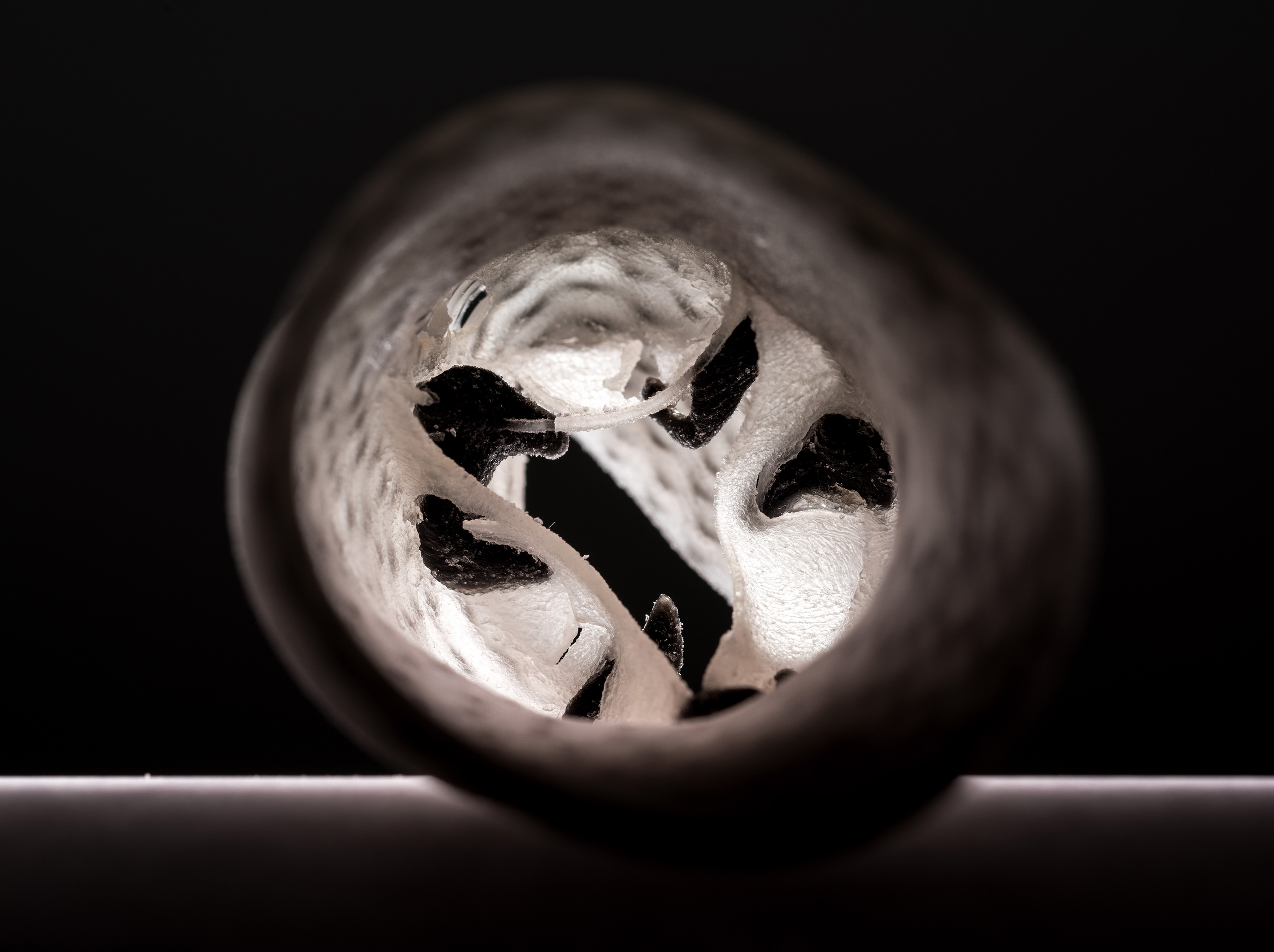
3-D Printed Heart Valve Model

ISyE Professor Chuck Zhang
For More Information Contact
Josh Brown
Research News
404.385.0500